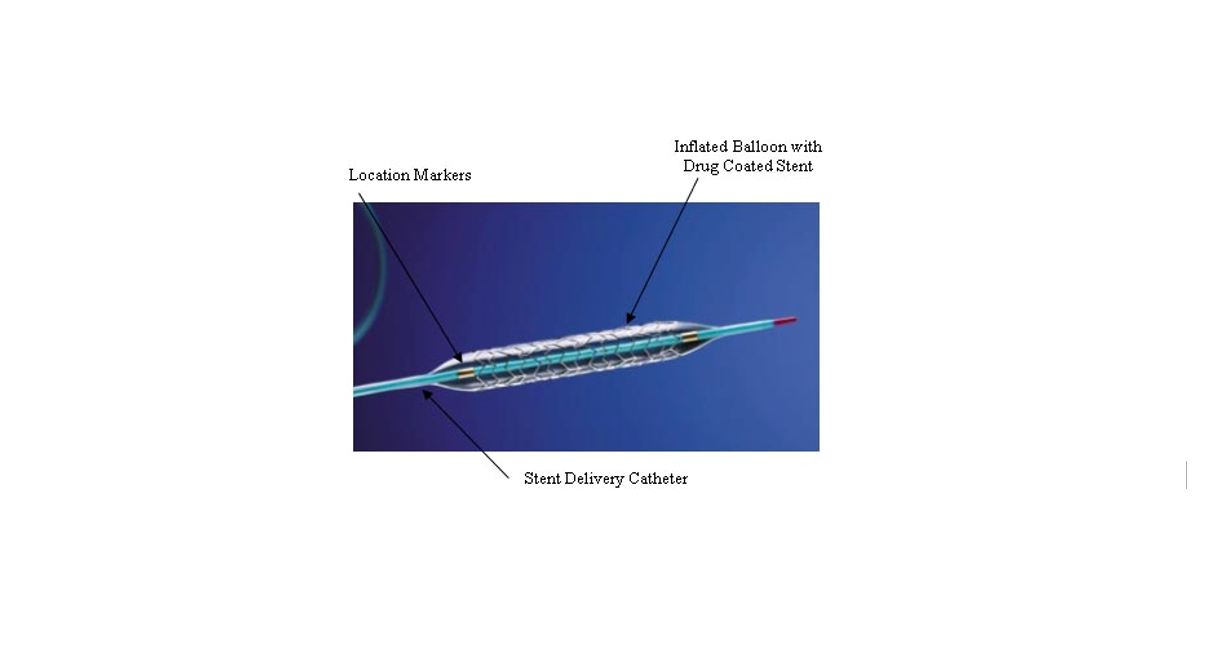
Coronary Stent
A coronary stent is a tube-shaped device placed in the coronary arteries that supply blood to the heart to keep the arteries open in patients suffering from coronary heart disease. The vast majority of stents used in modern interventional cardiology are drug-eluting stents (DES). They are used in a medical procedure called percutaneous coronary intervention (PCI). Coronary stents are divided into two broad types: drug-eluting and bare metal stents. As of 2023, drug-eluting stents were used in more than 90% of all PCI procedures.[1][2] Stents reduce angina (chest pain) and have been shown to improve survival and decrease adverse events after a patient has suffered a heart attack—medically termed an acute myocardial infarction.[3][4]
Similar stents and stenting procedures are used in atherosclerosis of arterial vessels of the limbs—particularly in the legs, such as in peripheral artery disease.[5]
Medical uses
Diagram of stent placement. In A, the catheter is inserted across the lesion. In B, the balloon is inflated, expanding the stent and compressing the plaque. In C, the catheter and deflated balloon have been removed. Before-and-after cross sections of the artery show the results of the stent placement.
Duration: 16 seconds.0:16
Arterial Stenting 3D Medical Animation
Cardiac stenting is achieved by PCI procedures in two distinct medical situations, when a patient has clearly suffered a heart attack and therefore PCI/stenting is being used in an emergency setting, termed 'primary PCI'. It is also a procedure used in patients that are exhibiting prolonged clinical symptoms of coronary artery narrowing (angina, evidence from stress test data, various imaging techniques etc.).[6]
Patients not undergoing primary PCI are usually awake during the placement of a coronary stent, though local anesthetics are used at the site of catheter entry, to ensure there is no pain. In reality practices vary, though patient comfort is a priority. Various techniques of pain management and anesthesia are practiced during current PCI stent placement procedures.[7]
The catheter/stent system is introduced into the body by penetrating a peripheral artery (an artery located in the arm or leg) and passed through the arterial system to deliver the DES into the blocked coronary artery. The stent is then expanded to dilate (open) blocked or narrowed coronary arteries (narrowed by plaque buildup), caused by a condition known as atherosclerosis. Peripheral arterial access is usually via the femoral (upper leg) or the radial artery (arm/wrist) and less commonly performed via the brachial or ulnar artery (wrist/arm). Historically, controlling bleeding at the point of arterial access after the procedure was an issue, modern arterial pressure bands and arterial closure system now exist which have helped control post procedure bleeding, but bleeding after the procedure is still a matter of concern.[8][9][10][11][12]
The 'stent tube mesh' is initially 'collapsed' onto the catheter, that catheter contains an inflating balloon component. In this collapsed state, it is small enough to be passed through 'relatively' narrow arteries and then inflated and compressed firmly against the diseased artery wall, by air pressure introduced via the still attached catheter, inflation time and pressure are recorded during this placement procedure. Consider an umbrella metaphor, initially unopened and then opened.[13][14]
Many significant treatment decisions are made in real time during the actual stent placement, the Interventional Cardiologist uses Intravascular ultrasound (IVUS) and fluoroscopic imaging data to assess the exact location, the true occlusion status. A radiopaque contrast dye is passed through the catheter and is used to visualize the arteries and evaluate the location of the narrowed vessel. This information is used in real time to decide how best to treat the occlusion(s). Information regarding the health and anatomy of the broader coronary blood supply can also be evaluated; as coronary vasculature varies from individual to individual. This data is captured on video and is valuable if any further treatments of a patient are necessary.[15][16][17]