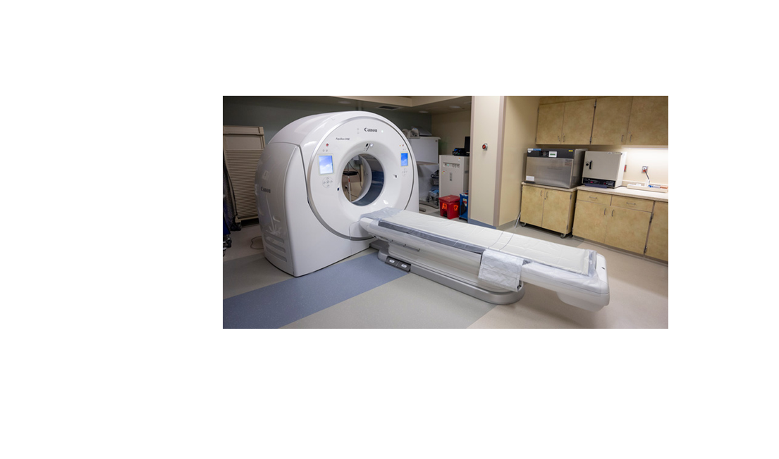
CT Scanner
A computed tomography (CT) scan, commonly referred to as a CT, is a radiological imaging study. The machine was developed by physicist Allan MacLeod Cormack and electrical engineer Godfrey Hounsfield.[1][2][3] Their development awarded them the Nobel Prize in Physiology or Medicine in 1979.[4] The first scanners were installed in 1974. Since then, technological advances and math have allowed single images to be computed into two-dimensional informative images.
The CT scan is essentially an X-ray study, where a series of rays are rotated around a specified body part, and computer-generated cross-sectional images are produced. The advantage of these tomographic images compared to conventional x-rays is that they contain detailed information about a specified area in cross-section, eliminating the superimposition of images, which provides a tremendous advantage over plain films. CT scans provide excellent clinicopathological correlation for a suspected illness.[5][6]
CT scan augments the physician's ability to diagnose a patient's illness accurately. Low-dose CT scans are proving useful in preventative medicine and cancer screening. The study was initially called a CAT scan representing computer axial tomography, where the table moved after each axial image was obtained.[7][8][9] In a spiral or helical scan, the table moves continuously as the x-ray source and detectors rotate. This reduces the duration of the study significantly to provide quick results in emergent situations. It rapidly substituted cerebral angiography for detecting head trauma injuries and brain masses in a fast and extremely reliable way.[10][11][12] A radiologic technician acquires CT scans, which are interpreted and reported by a trained radiologist.
Procedures
The CT scanner machine rotates the X-ray tube around the patient's body through a circular structure known as the gantry. Each time the machine rotates, computerized information is acquired. The patient is slowly moved up or down in the table, and different cross-section images are produced. In each rotation, a 2-dimensional image slice is constructed. Each subsequent image slice's thickness is decided on the operator's and the physician/radiologist's request but usually ranges from 1 to 10 millimeters. The gantry can be moved at the desired angle to accommodate the best cross-sectional image. When the desired number of slices is obtained, a scan is reproduced into the computer image and can easily be reproduced and stored.
The image is created using pixels according to its radiosensitivity and is displayed using the Hounsfield scale units, which are compared to known tissue density. Water is 0, air is negative 1000, and bone is positive 400 to 2000. Intravenous iodine can be injected into the bloodstream to demarcate blood vessels and tumors and identify infectious processes.[13] Intravenous iodine-based or oral barium-based contrast is used to visualize the digestive system. The images can be computer-tacked together to produce a 3-dimensional image of the area of interest. The CT scans are obtained in the cranial direction, meaning from feet to head. It is important to note that current CT machines display the image opposite the patient's side as the image is produced as viewed from the patient's foot. Thus, the right side of the image is the patient's left side.
Indications
A CT scan is used for numerous clinical indications depending on the organs to evaluate.[14] The CT scan can be utilized in both inpatient and outpatient clinical settings. In an emergent setting, it can rule out serious illness. Indications for obtaining a CT scan are to help the physician diagnose, narrow the differential diagnosis, and confirm the doctor's suspicions. This modality can also be used for cancer screening, staging, and follow-up; its use helps to perform biopsies adequately and to assist during a surgical procedure.
Brain: tumors, traumatic or spontaneous hematomas, stroke, edema, skull fracture, calcifications, arteriovenous malformations, hydrocephalus, sinusitis, and empyema
Neck: tumors, benign masses, thyroid nodules, lymphadenopathy
Chest: tumor, pneumonia, metastasis, benign masses, pulmonary edema, pleural edema, tuberculosis, pulmonary embolism, traumatic injury to the lungs, esophageal rupture, ingested foreign body, fibrosis
Abdomen: primary tumors, metastases, abscess, ascites, cholecystitis, appendicitis, renal calculi, pancreatitis, obstruction, lymphadenopathy, foreign body
Spine: fractures, degenerative changes, stability, osteomyelitis, disc pathology
Bone: complex bone fractures, eroded joints, knee, tumors, osteomyelitis
Gyn: cyst, fibromas, tumors
Screening: colon and lung cancer
CT colonography/colonoscopy is used to diagnose colon disease and early-stage cancer with good sensitivity and specificity.[15][16][17][18]
Low-emission CT can be used to diagnose lung cancer in smokers and former smokers with a high smoking history who are between 55 and 80 using a low radiation dose.[19][20][21][22]
Biopsy: CT guided to different organs for adequate tissue extraction
CT Angiography: brain, heart, lung, kidney, extremities
Intraoperative: Can be used for neuronavigation procedures during brain biopsy or tumor resection [23]